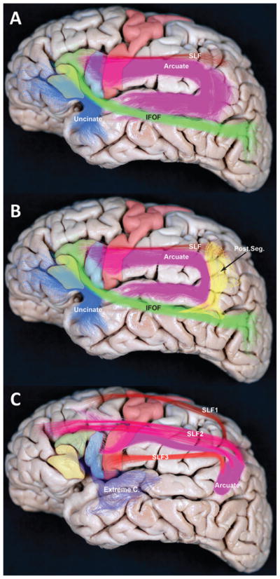
Fig. 4.
Subcortical pathways of the SLF and AF. A: This lateral view of a cadaveric brain shows the frontal operculum, along with several components of the SLF and AF. Solid shading indicates anatomical areas: pars orbitalis (yellow), pars triangularis (green), pars opercularis (blue), and motor cortex (red). Fiber pathways are indicated with colored bundles: SLF (red), AF (purple), inferior frontal-occipital fasciculus (IFOF; green), and uncinate fasciculus (blue). B: Areas and representative colors are the same as in the previous panel, but also shown is the posterior pathway (post. seg.) connecting posterior temporal and inferior parietal regions (yellow). C: SLF fascicles, as identified using autoradiography in the macaque monkey, are projected into the human brain. The SLF is believed to be divided into the 3 segments (SLF1, SLF2, and SLF3), along with the AF. Only SLF3 and the AF connect to the frontal operculum. Extreme C. = extreme capsule.

