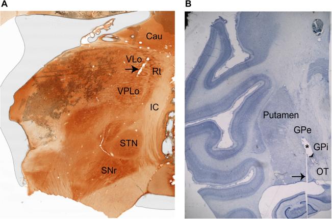
Figure 2.
Histological evidence for boundaries of VA/VLo and VPLo and DBS lead placement in the GPi. (A) Sagittal acetylcholinesterase stain shows the location of the thalamic sub-nuclei. The arrow indicates a recording track passing through the VLo. (B) Nissl stained coronal slice showing the location of the DBS lead (marked with an asterisk) passing through the pallidum. The histology image has been included here after obtaining permission from the publisher. The arrow points to an artifact in the slice preparation. The abbreviations are: Cau: caudate, IC: internal capsule, GPe: globus pallidus externus, GPi: globus pallidus internus, OT: optic tract, SNr: substantia nigra pars reticulate, STN: subthalamic nucleus, Rt: reticular thalamus, VLo: ventralis lateralis pars oralis, VPLo: ventralis posterior lateralis pars oralis.

