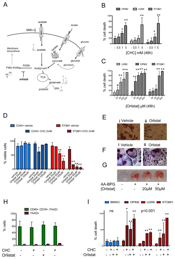
Figure 2. Pharmacological modulation of acetate metabolism leads to toxicity in MMC while sparing the surrounding BM resident cells.
A) Working model. Labeled acetate is imported from the extracellular environment through monocarboxylate transporters such as MCT1, is found inside cells (13C-NMR), where it may be activated to Acetyl-CoA. This can rapidly enter the TCA cycle and be transformed into glutamate (peaks 3 and 4) and participate in multiple reactions, including being incorporated into glutathione (GSH peak 5). Acetyl-CoA can also be a substrate for de-novo biosynthesis of fatty acids through the activity of fatty acid synthase (FASN), providing building blocks for cell membranes. CHC is a known inhibitor of MCT1, orlistat of FASN. Other monocarboxylic acids that can be involved in the same reactions include lactate, which is produced and released in the media by MMC and also pyruvate, as end-product of glycolysis. B) Treatment with α-cyano-4-hydroxycinnamic acid (CHC) induces MM cell death. Propidium Iodide staining and flow cytometry upon 48h incubation with 0–5mM CHC of OPM2, U266, or 5TGM1 cells (mean and standard deviation of 2–3 biological replicates per cell line, two-way ANOVA p<0.01 dose, cell line ns). C) Treatment with FASN inhibitor orlistat induces cell death in MMC lines. Propidium Iodide positive cells percentage by flow cytometry (mean and standard deviation of 2 to 8 biological replicates) upon treatment with 20–500μM orlistat in U266, OPM2, and 5TGM1 MMC for 48h (two-way ANOVA p<0.01). D) Cell viability by trypan blue staining, relative to untreated control, upon 48h treatment with orlistat (20–100 μM) plus vehicle (DMSO, open bars) or CHC 2mM (dashed bars) in primary mouse CD45+ spleen cells (blue bars) or 5TGM1 (red bars), showing sensitivity to single-agent and combined treatment in 5TGM1 (orlistat <0.01, interaction with CHC <0.05 by two-way ANOVA). E) Oil-red-O staining and hematoxylin counter-staining of ST2 cells induced to differentiate with adipogenic media (insulin, indomethacin, dexamethasone) for 14 days in the absence (left, i.) or presence (right, ii.) of 100 μM orlistat shows that long-term FASN inhibition prevents adipogenic differentiation but does not induce death of mesenchymal cells. F) Tartrate-resistant acid phosphatase (TRAP) staining of mouse bone marrow monocytes induced to differentiate to osteoclasts with M-CSF and RANKL shows formation of TRAP-positive multinucleated cells in the absence (left, i.) or presence (right, ii.) of 50 μM orlistat for 6 days. G) Alizarin-Red staining of mouse bone marrow stromal cells (BMSC) after standard culture (−) or osteogenic differentiation (+) with β-glycerophosphate (BPG) and ascorbic acid (AA) for 21 days shows mineralized matrix formation in the absence (−) or presence of orlistat (20 or 50 μM). H) Flow cytometry of mouse splenocytes treated for 48h with CHC (2mM) and/or Orlistat (100 μM) showing 7AAD+ dead cells (purple bars) vs. CD45+ CD19+ 7AA− B cells (ns). I) Cell death as % PI positive cells upon treatment with CHC and orlistat in primary BMSC (blue) vs. OPM2, U266 or 5TGM1 MMC (red) upon treatment with CHC, alone or in combination with Orlistat for 48h. Average of biological replicates, standard deviations as error bars, * p<0.05, ** p<0.001 (one-way ANOVA) relative to control.

