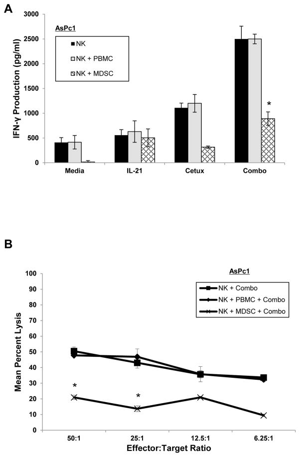
Figure 5. MDSCs inhibit NK cell IFN-γ production and ADCC.
Autologous PBMC-derived MDSCs or control PBMCs were co-cultured with healthy donor NK cells at a 0.5:1 ratio. In vitro generated MDSCs were found to be suppressive to T cell proliferation by CFSE assay (data not shown). (A) Purified IL-21-activated NK cells were co-cultured with cetuximab-coated KRAS mutant AsPc1 cancer cells alone or in the presence of autologous PBMCs or MDSCs. Control conditions consisted of medium alone, IL-21 alone, or cetuximab alone. Culture supernatants were harvested at 48 hrs and analyzed for IFN-γ by ELISA. (B) Purified human NK cells were incubated overnight in medium alone in medium containing 10 ng/ml of IL-21. The lytic activity was tested against cetuximab-coated KRAS mutant AsPc1 cancer cells in the presence of autologous PBMCs or MDSCs in a standard 4 hr chromium release assay. Each graph depicts the results from one representative donor ± SD. Three normal donors were tested per cell line. The asterisk (*) denotes p<0.001 versus all conditions shown.

