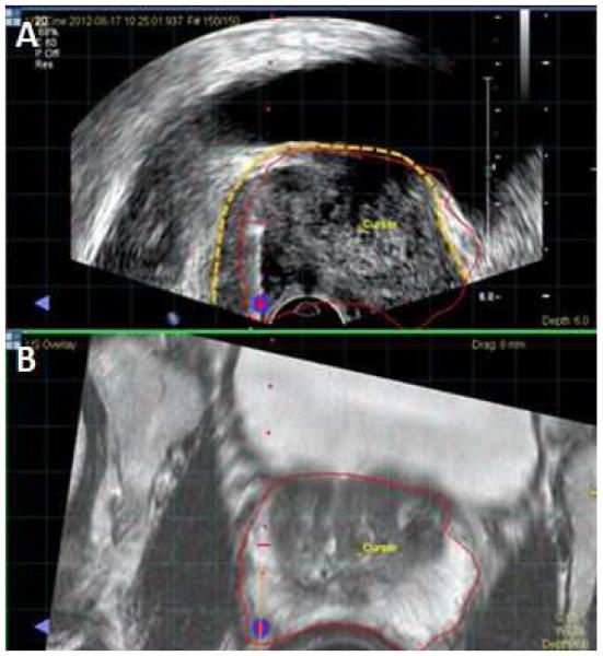
Figure 2.
Screen Capture from fusion biopsy platform. A. Fused image with MR prostate segmentation (red line) overlaid on top of ultrasound. The ultrasound prostate border is highlighted by the dotted yellow line. The MR segmentation does not perfectly align with the ultrasound prostate border, illustrating the presence of registration error. B. T2 weighted MRI with prostate border segmented in red.

