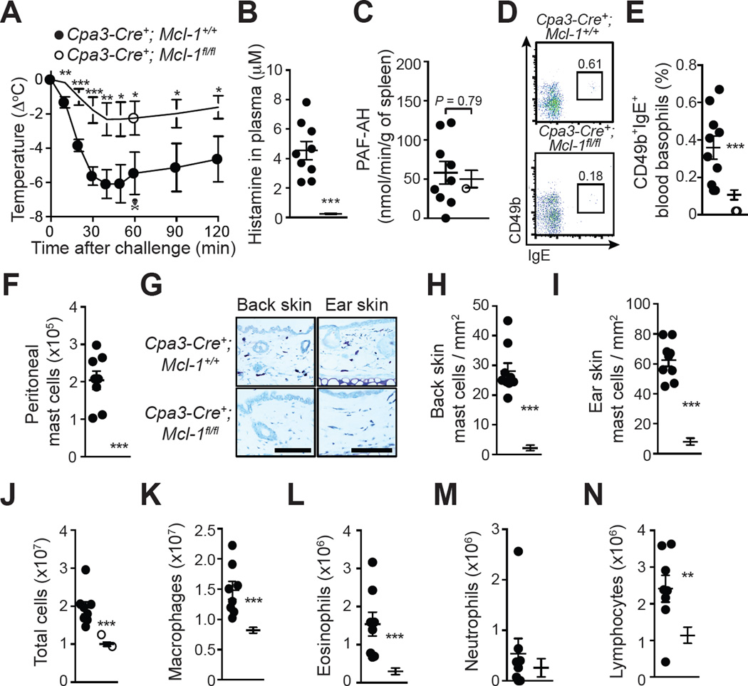
Figure 5. Assessment of OVA-induced ASA in genetically MC-deficient and basophil-depleted Cpa3-Cre; Mcl-1fl/fl mice.
(A) OVA-induced hypothermia in OVA-sensitized Cpa3-Cre+; Mcl-1+/+ and Cpa3-Cre+; Mcl-1fl/fl mice. (B) Levels of histamine in the plasma 20 min after challenge. (C) PAF-AH activity in the spleen 20 min after challenge. (D–E) Representative FACS profile (D) and percentage (E) of blood basophils (CD49b+; IgE+) 24 h before challenge. (F) Numbers of MCs in the PLF 3 days after challenge. (G–I) Toluidine blue staining for MCs (G) and MC numbers (H, I) in sections of back skin and ear pinna. (J-N) Numbers of leukocytes in the PLF 3 days after challenge. Data are pooled from three independent experiments (total n=9–14/group). *, ** or *** = P < 0.05, 0.01 or 0.001. The crossbones symbol indicates death of one mouse. Scale bars: 100 µm.

