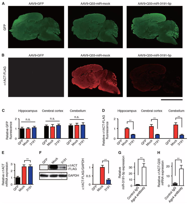
Fig. 6. AAV9-mediated therapeutic delivery of miR-3191-5p blocks CACNA1A IRES–driven α1ACT translation in mice.
(A and B) Representative immunofluorescence images of AAV9-injected mouse brain and cerebellum from 4-week-old mice stained with GFP-specific (A) and FLAG-specific (B) antibodies and fluorescent secondary antibody. Scale bar, 5 mm. (C and D) Relative GFP (C) and α1ACT-FLAG (D) immunofluorescence intensities in AAV9-injected mouse hippocampus, cerebral cortex, and cerebellum (n = 6). Relative GFP and relative FLAG immunofluorescence are expressed as signal intensities per unit area quantified by NIH ImageJ software. (E) Relative α1ACT mRNA expression in the cerebellum of AAV9-injected mice at 4 weeks of age (n = 4). (F) Western blot and densitometric analyses showing α1ACT-Q33-FLAG expression in the cerebellum of AAV9-injected mice at 4 weeks of age (n = 4). (G and H) Amount of miR-3191-5p (G) and α1ACT-Q33 (H) mRNA binding to Ago4 that was immunoprecipitated using an antibody against Ago4 or a control IgG in the cerebellum of AAV9–Q33–miR-3191-5p mice at 4 weeks of age (n = 6). GFP, AAV9-GFP mice; mock, AAV9–Q33–miR-mock mice; 3191, AAV9–Q33–miR-3191-5p mice. All data represent means ± SEM. **P < 0.01. One-way ANOVA in (C). Student’s t test in (D) to (H).

