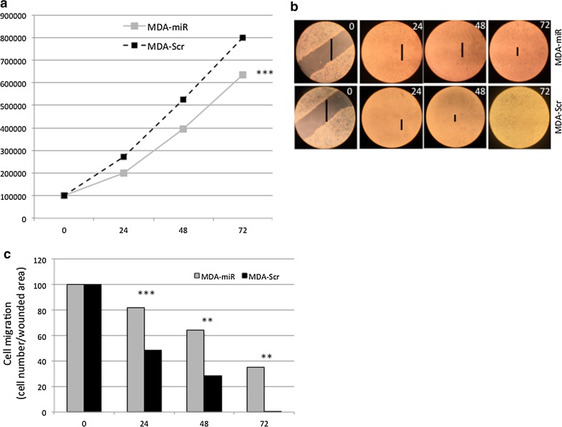
Fig. 4.
a Growth curve of MDA-Scr (dashed black line) and MDA-miR (solid gray line) cell lines. All data are presented as mean ± SD of experiments for each point at each time point (***t test p value = 1.28 × 10⁻⁷, n = 27). b Behavior of MDA-Scr and MDA-miR cells in wound-healing test. c ImageJ quantification of the effect of the miR-567 overexpression in MDA-miR compared to MDA-Scr cells, on wound-healing assay. All data are presented as mean ± SD of experiments for each bar at each time point (**p value < 0.01, ***t test p value < 0.001, n = 3, respectively)

