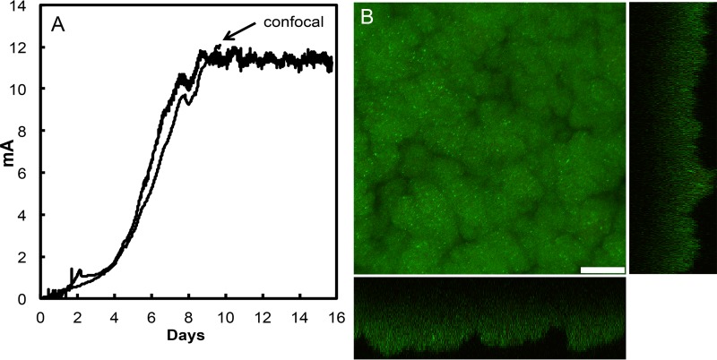
FIG 2 .
Growth of G. sulfurreducens strain MP on graphite anodes. (A) Time course of current production in duplicate cultures. One anode was removed for imaging with confocal scanning laser microscopy at the time designated. (B) Confocal scanning laser micrographs of strain MP anode biofilms harvested on day 10 (indicated in panel A). Top-down three-dimensional, lateral side views (right image) and horizontal side views (bottom image) show cells stained with LIVE/DEAD BacLight viability stain. Bar, 25 µm.

