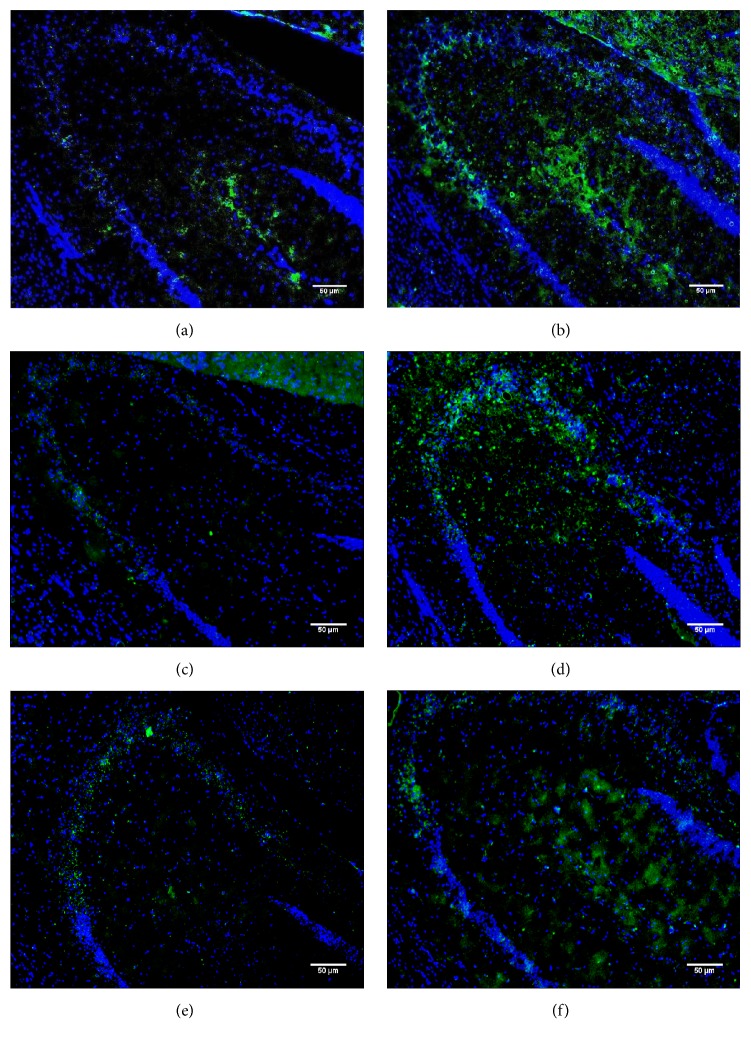
Figure 6.
Hippocampal endothelin-1 distribution in 3-, 6-, and 12-month-old wild-type mice and their CathAS190A littermates. Images show the hippocampus of wild-type (a, c, and e) and CathAS190A (b, d, and f) mice after immunostaining with endothelin-1 antibody (green) and DAPI (blue). Images were captured at 20x magnification.

