
Figure 1.
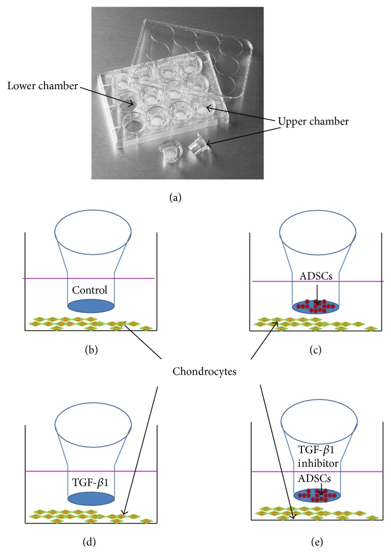
The experimental model. (a) The picture of transwell chamber. ((b)–(e)) The schematic diagram of the four groups, respectively, for chondrocytes cultured in normal culture medium (b), chondrocytes cocultured with ADSCs together (c), chondrocytes cultured with TGF-β1-containing medium (d), and chondrocytes cocultured with both ADSCs and TGF-β1 inhibitor (e).