Fig. 2.
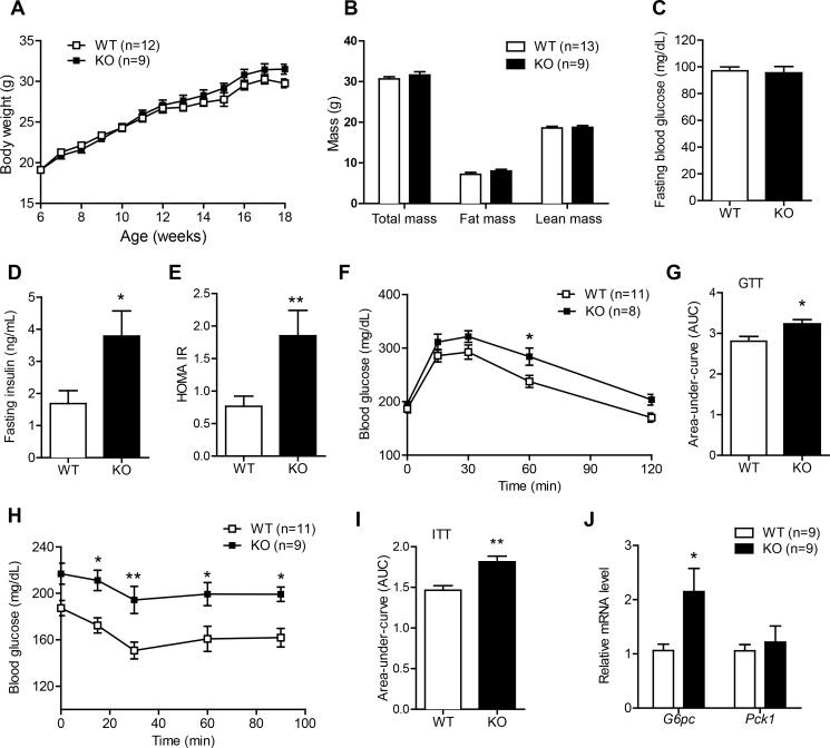
Ctrp1 KO mice fed a LFD develop insulin resistance. A: body weight gain over time of WT (n = 12) and KO (n = 9) male mice fed a control LFD. B: body composition analysis of lean and fat mass of WT (n = 13) and KO (n = 9) male mice. C–E: overnight fasting blood glucose (C), insulin (D), and homeostatic model assessment of insulin resistance (HOMA-IR) index (E) of WT (n = 10) and KO (n = 8) male mice. F: blood glucose levels of WT (n = 11) and KO (n = 8) male mice subjected to a glucose tolerance test (GTT). Glucose was delivered by an intraperitoneal injection. G: area under the curve (AUC) for the GTT data shown in F. H: blood glucose levels of WT (n = 11) and KO (n = 9) male mice subjected to an insulin tolerance test (ITT). I: AUC for the ITT data shown in H. J: quantitative real-time PCR analysis of gluconeogenic gene [glucose 6-phosphatase (G6Pc) and phosphoenolpyruvate carboxykinase 1 (Pck1)] expression in the liver of WT (n = 9) and KO (n = 9) male mice. Expression levels were normalized to β-actin. All data are expressed as means ± SE. *P < 0.05; **P < 0.01.

