Fig. 3.
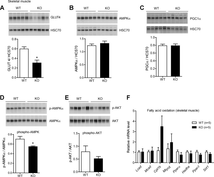
Reduced skeletal muscle glucose transporter (GLUT)4 and AMP-activated protein kinase (AMPK)α levels in Ctrp1 KO mice fed a LFD. A–C: quantitative Western blot analysis of skeletal muscle GLUT4 (A), AMPKα (B), peroxisome proliferator-activated receptor (PPAR)-γ coactivator-1α (PGC1α; C) levels in WT (n = 5) and KO (n = 5) male mice. Data were normalized to HSC70. D and E: quantitative Western blot analysis of phosphorylated (p)AMPKα (Thr172; D) and pAKT (Ser473; E). Data were normalized to total AMPKα or AKT. The total AMPKα blot shown in B is the same as shown in D. F: expression of genes involved in skeletal muscle fat oxidation. Expression levels were normalized to 18S rRNA. Lcad, long-chain acyl-CoA dehydrogenase; Mcad, medium-chain CoA dehydrogenase; Cpt, carnitine palmitoyltransferase; Mlycd, malonyl-CoA decarboxylase; Hadha, hydroxyacyl-CoA dehydrogenase/3-ketoacyl-CoA thiolase/enoyl-CoA hydratase (Trifunctional Protein) α-subunit; Sirt1, sirtuin 1. All data are expressed as means ± SE. *P < 0.05.

