Fig. 4.
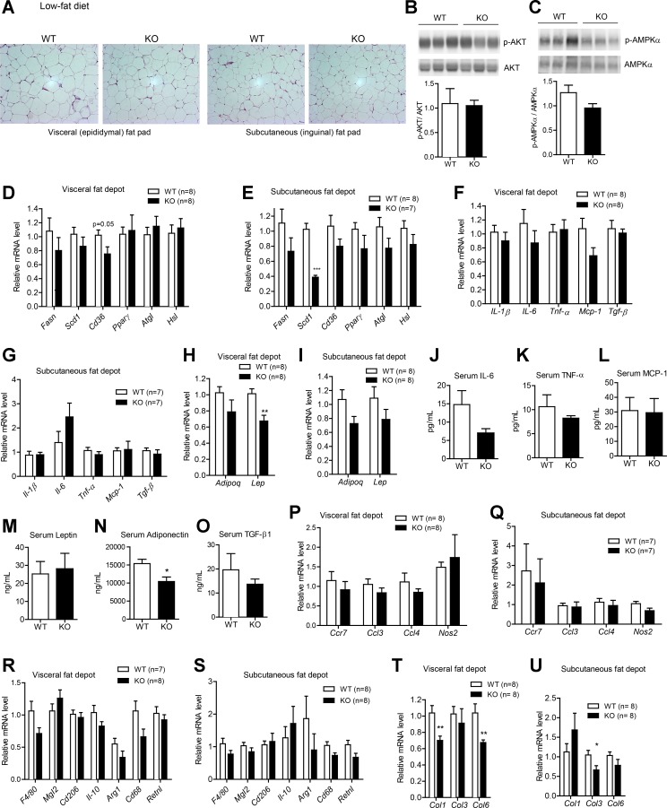
Metabolic, inflammatory, and fibrotic gene expression in the adipose tissue of Ctrp1 KO male mice fed a LFD. A: representative histology (hematoxylin and eosin stain; magnification: ×200) of the visceral (epididymal) fat pad of KO male mice and WT littermate controls. B and C: quantitative Western blot analysis of pAKT (B) and pAMPKα (C) in visceral adipose tissue of WT (n = 6) and KO (n = 6) mice. D and E: expression of lipid uptake (Cd36), synthesis [fatty acid synthase (Fasn), stearoyl-CoA desaturase-1 (Scd1), and PPAR-γ] and lipolysis [adipose triglyceride lipase (Atgl) and hormone-sensitive lipase (Hsl)] in the visceral (epididymal; D) and subcutaneous (inguinal; E) fat depot of WT (n = 8) and KO (n = 7–8) mice. F and G: expression of inflammatory [IL-1β, IL-6, TNF-α, and monocyte chemotactic protein (Mcp)-1] and profibrotic [transforming growth factor (TGF)-β1] cytokine genes in the visceral (epididymal; F) and subcutaneous (inguinal; G) fat depot of WT (n = 7–8) and KO (n = 7–8) mice. H and I: expression of adiponectin (Adipoq) and leptin (Lep) in the visceral (epididymal; H) and subcutaneous (inguinal; I) fat depot of WT (n = 8) and KO (n = 8) mice. J–M: serum levels of IL-6 (J), TNF-α (K), MCP-1 (L), leptin (M), adiponectin (N), and TGF-β (O) in WT (n = 10) and KO (n = 8) mice. P and Q: expression of proinflammatory M1 macrophage marker genes in the visceral (epididymal; P) and subcutaneous (inguinal; Q) fat depot of WT (n = 7–8) and KO (n = 7–8) mice. Ccr, chemokine (C-C motif) receptor; Ccl, chemokine (C-C motif) ligand; Nos, nitric oxide synthase. R and S: expression of anti-inflammatory M2 macrophage marker genes in the visceral (epididymal; R) and subcutaneous (inguinal; S) fat depot of WT (n = 7–8) and KO (n = 8) mice. Mgl2, macrophage galactose N-acetyl-galactosamine-specific lectin 2; Arg, arginase; Retn, resistin. T and U: expression of fibrotic collagen (Col) genes in the visceral (epididymal; T) and subcutaneous (inguinal; U) fat depot of WT (n = 8) and KO (n = 8) mice. Expression levels were normalized to 36B4 [also known as ribosomal phosphoprotein P0 (RPLP0)]. All data are expressed as means ± SE. *P < 0.05; **P < 0.01; ***P < 0.005.

