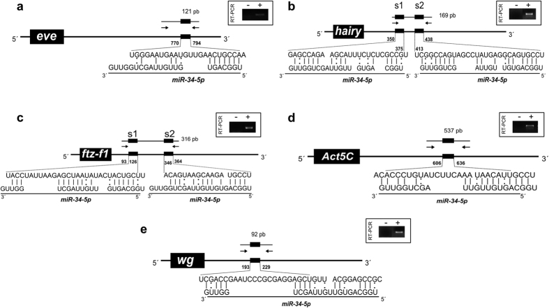
Figure 4. Predicted binding-sites in the developmental genes 3′UTRs for miR-34-5p by RNAhybrid.
Schematic representation of honey bee miR-34-5p binding-sites and RT-PCR amplified 3′UTRs of validated genes in the dual-luciferase assay. The box in the upper-right region of each scheme contains the amplified regions and the negative control of the RT-PCR reactions. The nucleotide positions of miR-34-5p binding sites (small black box), numbered from the stop codon, are shown on 3′UTR feature. The arrows indicate 3′UTR regions that were PCR amplified. Perfect matches are indicated by a line, and G:U pairs by a colon.

