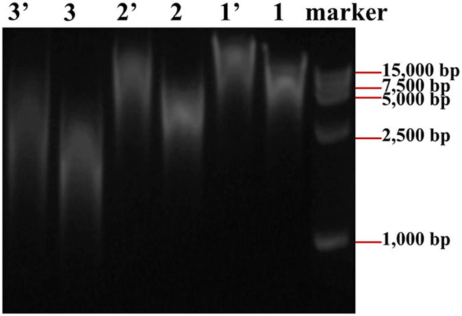
Figure 6. Gel images of the DNAs before and after ligation reaction with 1 and 1′- fragmented genomic DNAs at 0.10 MPa before and after ligation, 2 and 2′- fragmented genomic DNAs at 0.20 MPa before and after ligation, 3 and 3′- fragmented genomic DNAs at 0.30 MPa) before and after ligation.

The bubbling time is 60 min for all samples. Molecular weight marker DL 15,000 was used to measure the size of the DNAs.
