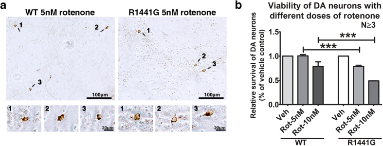
Figure 2. Cell viability of LRRK2R1441G mutant mouse primary dopaminergic (DA) neurons after rotenone exposure.
Primary mesencephalic DA neuronal cultures (DIV7) from mutant and WT mouse embryos (E14.5) were treated with 5 or 10 nM rotenone (Rot), or vehicle (Veh: DMSO; 0.01% in culture) for 48 hr. (a) Immunostaining of DA neurons using anti-tyrosine hydroxylase (TH) antibody after treated with 5 nM rotenone. Total number of TH-positive cells in culture was counted by two blinded observers independently under light microscope. (b) Relative survival of mutant DA neurons was significantly lower after exposure to rotenone than similarly treated WT groups. Data represents mean ± standard error of mean (SEM) from three independent experiments (N ≥ 3). Statistical significance between groups was analyzed by one-way ANOVA and Tukey post-hoc test, ***P < 0.001.

