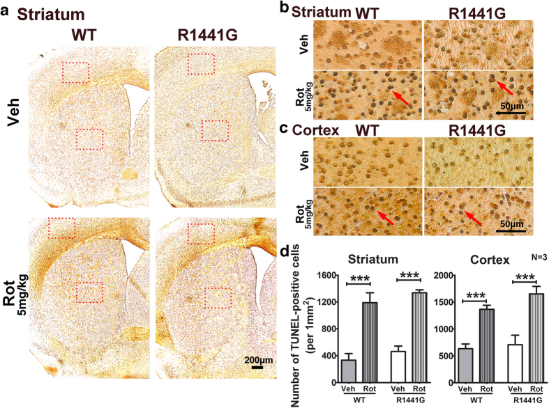
Figure 7. Number of apoptotic nuclei in striatum and cortex of WT and LRRK2R1441G mutant mice after 50-week oral rotenone administration.
(a) Low magnification (X40) and (b,c) high magnification (X400) photomicrographs in striatal and cortical regions after TUNEL assay. TUNEL-positive nuclei (red arrow) were seen in striatum and cortex of both WT and mutant rotenone-treated mice. N = 3. Scale = 200 μm (a) and 50 μm (b,c). (d) Quantification of TUNEL-positive cells in striatum and cortex (rectangular box). Statistical analysis of TUNEL-positive cell numbers in stained striatum and cortex regions showed that rotenone induced similar levels of apoptosis in both WT and mutant mice. Data represents mean ± standard error of mean (SEM) from three mice per group; Statistical significance between groups was analyzed using Student’s unpaired t-test, ***P < 0.001.

