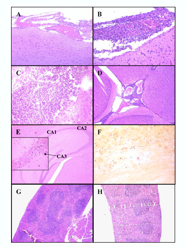
Figure 3.

Histological analysis of the brain and spleen of mice infected with type 4 S. pneumoniae. MF1 outbred mice were infected by the i.c. subarachnoidal route with the TIGR4 strain and humanely killed before reaching the moribund state. Brains (A-F) and spleens (G-H) were excised, fixed in formalin, embedded in paraffin, and stained with either haematoxilin-eosin or Gram. A. Mild inflammatory changes with congested leptomeningeal blood vessels and PMNs margination. B-C. Severe inflammation characterised by cellular exudates composed of PMNs entrapped in fibrin in the subarachnoid space. In panel C, the fibrin web is clearly visible. D. Acute inflammation in the ventricular spaces. E. Brain damage in the hippocampus: neuronal shrinkage in the CA3 hippocampal region is shown in the inset. The location of CA1, CA2 and CA3 areas is represented. F. Gram staining of pneumococci in the subarachnoid space of the brain of moribund mice. Short chains (mainly diplococci) of Gram positive bacteria surrounded by granulocytes. G-H. Haematoxilin-eosin stained spleen sections. A distinct congestion of the red pulp together with considerable modifications of the white pulp are present in the spleen of animals infected with S. pneumoniae (G) compared to controls (H).
