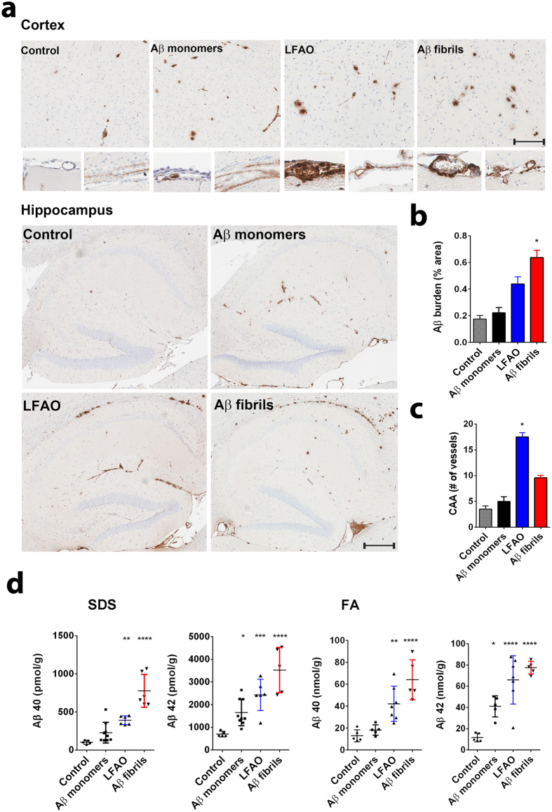
Figure 2. LFAOs selectively induce widespread amyloid deposition and CAA in TgCRND8 mice.
(a) Newborn CRND8 mice were injected with 4 μL Aβ fibrils, LFAO, Aβ monomers (10 μM) in the cerebral ventricles. Control group received PBS. 3 months later brains were extracted and one hemibrain fixed and stained with anti-pan- Aβ mAb5Biotin antibody. Amyloid staining of plaques and CAA in the representative paraffin sections is shown in the cortex, meningeal vessels and choroid plexus and in the hippocampus of injected mice. Scale Bar, 500 μm (cortex and CAA), 250 μm (hippocampus). (b) Quantification of Aβ positive immunostaining shows significantly increased amyloid plaque burden (immunostained with anti-pan- Aβ mAb5Biotin) Aβ fibrils injected mice compared to control mice. Data represents mean ± sem. n = 6–10/group. ***p < 0.01, unpaired two-tailed t test. (c) Quantification of CAA. Aβ positive positive blood vessels in the meningies and throughout the brain tissue were evaluated in a blind manner and given a qualitative score from 0 to 3. Vessels with scores 2 or 3 were counted. LFAO injected mice have significantly higher levels of CAA compared to control. Data represents mean ± sem. n = 6–10/group. ***p < 0.01, unpaired two-tailed t test. (d) Biochemical analyses of sequentially extracted Aβ42 and Aβ40 levels by end-specific sandwich ELISA show significantly increased SDS soluble and formic acid extractable insoluble Aβ levels in LFAO and Aβ fibrils injected mice compared to control mice. Small increase in Aβ levels was also detected in Aβ monomer injected mice. Data represents mean ± sem. n = 6–10 mice/group. N = 6–10, *p < 0.05, **p < 0.01, ***p < 0.001, ****p < 0.0001, 2 way Anova with Tukey’s multiple comparison test).

