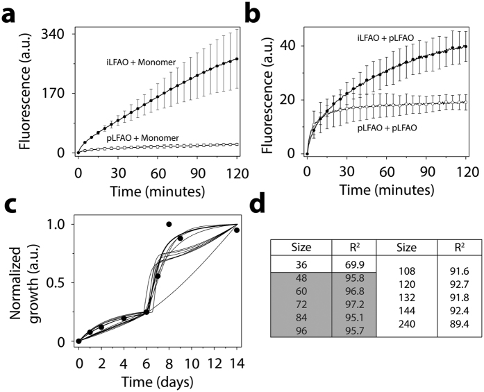
Figure 7. Identification of the propagating unit within sLFAO fibrils.
(a) Aβ monomer (30 μM) seeded with 6 μM of either iLFAOs (●) or pLFAOs (○). (b) pLFAOs (10 μM) seeded with 2 μM of either iLFAOs (●) or pLFAOs (○). For both a and b, kinetic measurements were taken every 30 s beginning immediately after the addition of 10 μM ThT. (c) Numerical simulations of sLFAO aggregate formation (●, Fig. 3a), along with (d) R2 values for each respectively LFAO assembly (gateway) considered.

