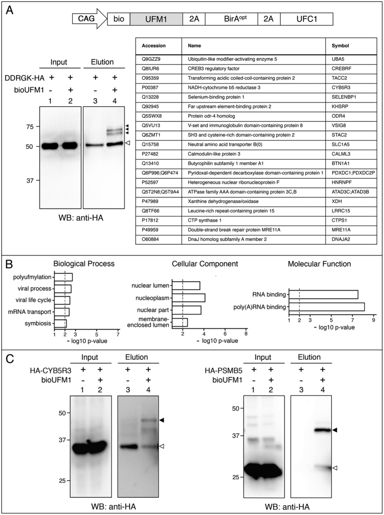
Figure 6. Isolation and identification of bioUFM1-conjugates in mammalian cells.
(A) Up: schematic representation of the bioUFM1 vector for mammalian cells. Below, left: Validation of HA-DDRGK. Right, top twenty bioUFM1-modified proteins. (B) GO analysis for biological process, cellular component and molecular function of the selected bioUFM1-conjugated protein set (n = 82). (C) Validation of bioUFM1-modified proteins CYB5R3 and PSMB5 fused to HA tag. In the elution panels (lanes 3 and 4), black arrowheads indicate the modified forms of the respective proteins. Residual non-specific interactions of non-modified forms are indicated by white arrowheads. Molecular weight markers are shown to the left.

