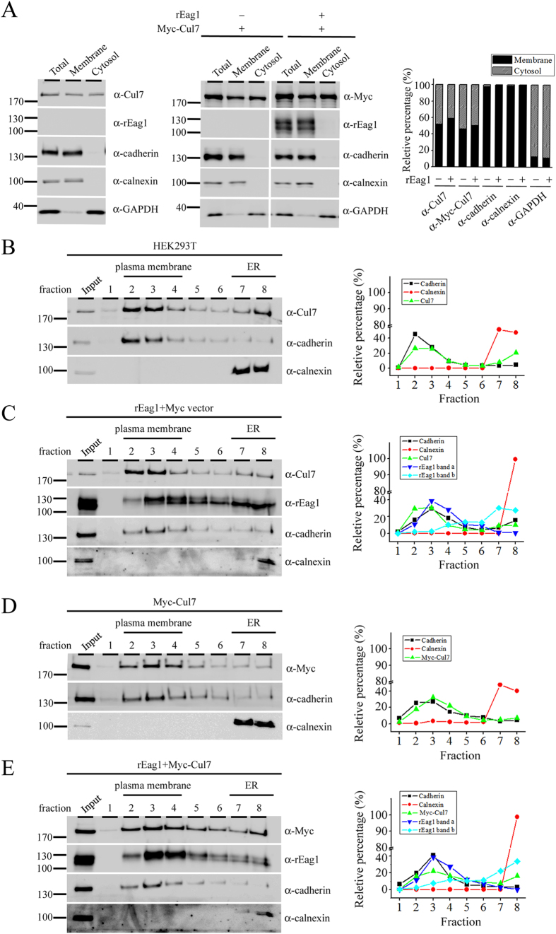
Figure 6. Centrifugation and fractionation analyses support the localization of Cul7 at the plasma membrane.
HEK293T cell homogenates were analyzed by differential centrifugation (A) or sucrose gradient fractionation (B–E). In both cases, total homogenates (Total) were ultracentrifuged and thereafter separated into the supernatant (Cytosol) and the pellet (Membrane) fractions. Three endogenous proteins in HEK293T cells were used as specific markers for distinct subcellular compartments: cadherin (plasma membrane protein), calnexin (ER-resident membrane-associated protein), and GAPDH (cytosolic protein). (A) In differential centrifugation analyses, both endogenous Cul7 (detected by the anti-Cul7 antibody) and over-expressed Myc-Cul7 (detected by the anti-Myc antibody) were found to be present in the cytosol as well as the membrane fractions. In contrast, rEag1 was present in the membrane fraction only. Shown to the right is the quantification of the relative percentage of membrane and cytosol distributions (with respect to the total signal) for each protein (n = 3). (B–E) In sucrose gradient fractionation analyses, the membrane pellet fraction (Input) was sedimented through a discontinuous sucrose gradient and further divided into 8 fractions. Fractions 2–4, wherein cadherin was detected, correspond to the plasma membrane fractions. Fractions 7–8, wherein calnexin was detected, indicate the ER membrane fractions. Shown to the right is the densitometric quantification of each representative immunoblot that highlights the relative distribution (with respect to the total signal) in different fractions for a specific protein. (B) Endogenous Cul7 in HEK293T cells was predominantly found in the plasma membrane fractions, and to a lesser extent in the ER membrane fractions. (C) rEag1 protein bands a and b were preferentially detected in the plasma membrane and the ER membrane fractions, respectively. (D) A significant portion of over-expressed Myc-Cul7 was localized in the plasma membrane fractions. (E) Myc-Cul7 and rEag1 protein band a co-localized in the plasma membrane fractions. The gels were run under the same experimental conditions. Uncropped images of immunoblots are shown in Supplementary Figure S8.

