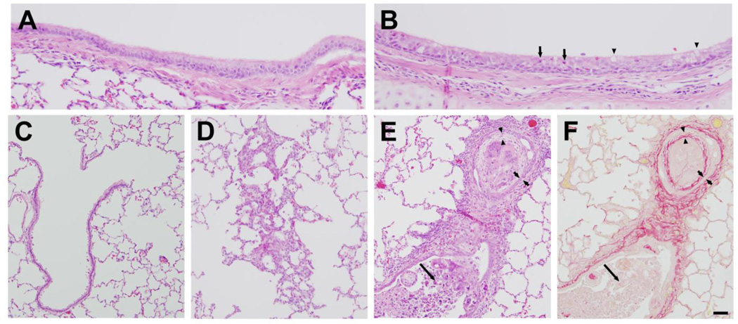
Figure 11. Lung histology 7 days after exposure.
A) Lobar bronchus from air-exposed rabbit showing normal airway structure. B) Lobar bronchus from chlorine-exposed rabbit showing repaired pseudostratified epithelium containing granulocytes (arrows) and increased goblet cells (arrowheads). C) Section from air-exposed rabbit showing normal structure of terminal bronchiole and alveoli. D) Hyperplasia and abnormal alveolar structure in chlorine-exposed rabbit. E) Terminal bronchiole from chlorine-exposed rabbit containing inflammation and bronchiolitis obliterans lesion. Part of the airway contains an identifiable lumen with considerable inflammatory cells and cell debris (long arrow). Part of the airway cut in cross-section contains concentric layers of abnormal epithelium (bracketed by short arrows), fibrosis (bracketed by arrowheads), and inflammatory cells and cell debris filling the lumen. F) Adjacent section to panel E stained with Sirius red. Red staining shows normal collagen in airway wall and abnormal fibrotic layer (bracketed by arrowheads) inside the epithelium (bracketed by short arrows). Long arrow indicates inflammation and cell debris inside airway lumen. Scale bar represents 25 µm for panels A and B, 50 µm for panels C–F. Results are representative of 9 chlorine-exposed and 7 sham-exposed rabbits.

