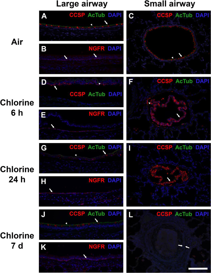
Figure 12. Immunohistochemical staining for airway epithelial cell markers.
A) Large airway from air-exposed rabbit showing normal distribution of ciliated cells (green AcTub staining indicated by arrowhead) and club cells (red CCSP staining indicated by arrow). B) Large airway from air-exposed rabbit showing normal distribution of basal cells (red NGFR staining indicated by arrows). C) Small airway from air-exposed rabbit showing normal distribution of ciliated cells (green AcTub staining indicated by arrowhead) and club cells (red CCSP staining indicated by arrow). D) Large airway from rabbit 6 h after exposure to 800 ppm chlorine for 4 min. AcTub (green, arrowhead) and CCSP (red, arrow) staining is confined to epithelial cell debris that has sloughed from the airway. E) Large airway from rabbit 6 h after exposure to 800 ppm chlorine for 4 min. Surviving basal cells staining for NGFR remain attached to the airway wall and adopt a more spread morphology (arrow). F) Small airway from rabbit 6 h after exposure to 800 ppm chlorine for 4 min. Most CCSP staining is observed in injured epithelium that has sloughed from the airway (arrow). In some airways, a few residual club cells remain attached (arrowhead). AcTub staining is virtually absent from most small airways. G) Large airway from rabbit 24 h after exposure to 800 ppm chlorine for 4 min. Similar to what was observed at 6 h, AcTub (arrowhead) and CCSP (arrow) staining is confined to sloughed epithelial debris in the airway lumen and is not present in the airway proper. H) Large airway from rabbit 24 h after exposure to 800 ppm chlorine for 4 min. NGFR-stained basal cells with squamous morphology are observed in the airway. I) Small airway from rabbit 24 h after exposure to 800 ppm chlorine for 4 min. Most CCSP staining is observed in injured epithelium that has sloughed from the airway (arrow). Lack of DAPI staining suggests that this cellular debris has continued to break down between 6 and 24 h. J) Large airway from rabbit 7 days after exposure to 400 ppm chlorine for 8 min. A regenerated airway epithelium containing AcTub-stained ciliated cells (arrowhead) and CCSP-stained club cells (arrow) is present, but it appears to have a lower density of club cells compared with air-exposed mice. K) Large airway from rabbit 7 days after exposure to 400 pm chlorine for 8 min showing normal morphology of NGFR-stained basal cells (arrow). L) Small airway with bronchiolitis obliterans lesion from rabbit 7 days after exposure to 400 ppm chlorine for 8 min. The epithelial layer lining this airway (bracketed by arrows) is devoid of staining for AcTub and CCSP. Scale bar represents 100 µm for all panels. Results are representative of 7 rabbits/group for A–C, 6 rabbits/group for D–F, 9 rabbits/group for G–I, and 9 rabbits/group for J–L.

