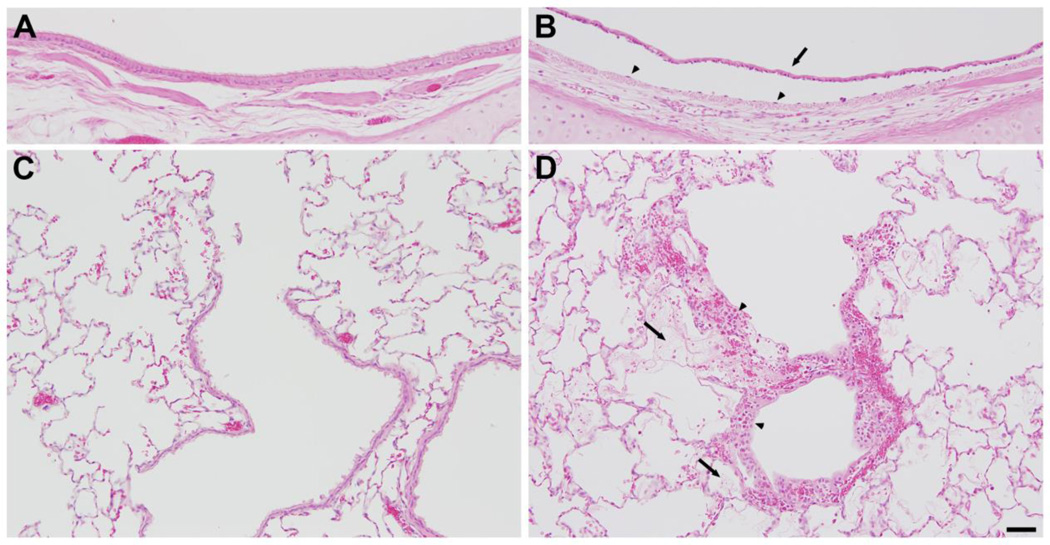
Figure 4. Lung histology 6 h after exposure to 800 ppm chlorine for 4 min.
All panels show hematoxylin and eosin staining of lung sections collected 6 h after exposure to 800 ppm chlorine for 4 min. A) Lobar bronchus from air-exposed rabbit showing normal airway structure. B) Lobar bronchus from chlorine-exposed rabbit showing sloughed epithelium (arrow) and residual, thinly spread basal cells (arrowheads). C) Section from air-exposed rabbit showing normal structure of alveoli and terminal bronchiole. D) Section from chlorine-exposed rabbit showing injured terminal bronchiole, including damaged epithelium and inflammation (arrowheads). Alveolar injury with fibrin deposition is indicated by arrows. Scale bar in D represents 50 µm for all panels. Results are representative of 8 chlorine-exposed and 8 sham-exposed rabbits.

