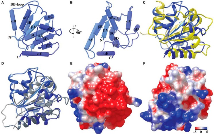
FIGURE 1.
Structure of the TIR domain of human BCAP. A and B, the BCAP TIR domain has the typical TIR fold consisting of five parallel β-strands βA–βE) surrounded by five helices (αA–αE). C and D, structural superposition of BCAP-TIR and the TIR domain of TLR2 (C) and MAL/TIRAP (D). Electrostatic surface potential of BCAP-TIR (E and F) using the orientations shown in panels A and B is colored from −5 (red) to +5 kcal/mol (blue). Electrostatic potentials were calculated using Chimera.

