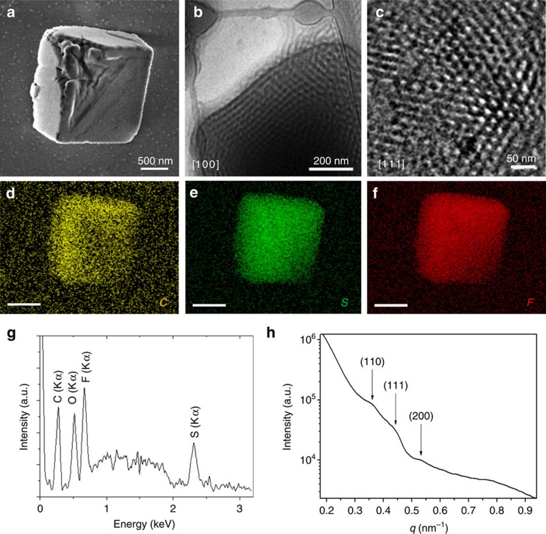
Figure 3. Triply periodic structures of cubosomes.
The electron micrographs of the self-assembled aggregates of PAA45-b-PIL23 in water/THF solution recorded 2 days after adding water and aging the dispersion at 21 °C. Self-assembling conditions: 2 mg ml−1 THF solution of PAA45-b-PIL23, water/THF volume ratio (R)=1.6. (a) SEM micrograph of the dried cubosomes after dialysis. (b) Cryo-TEM image of the [100] facet of a cuboid particle. (c) Enlarged surface image recorded in the central area of a cuboid particle from the [111] facet. (d–f) Elemental mappings (scale bar: 100 nm) show the uniform distribution of the elements C, S and F throughout the cuboid structure. The homogeneous distribution is in agreement with the interpenetrating bicontinuous structure expected for cubosomes. (g) Energy-dispersive X-ray spectrum. A Si peak was subtracted that originated from the silicon wafer on which the cubosomes were deposited. (h) SAXS diagram indicates a double diamond (Pn3m) lattice with a=23.5 nm (q/q* ∼ ).
).

