Figure 1.

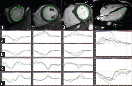
CMR-FT postprocessing. Short-axis apical (1), midventricular (2), basal (3) and 4-chamber long-axis (4) views with relevant endocardial contour drawn in a KD patient. Radial strain (a) and strain rate (b), circumferential strain (c) and strain rate (d), and longitudinal strain and strain rate (4, mid and lower row, respectively) results are provided below each slice. CMR-FT: Cardiac magnetic resonance feature tracking; KD: Kawasaki disease
