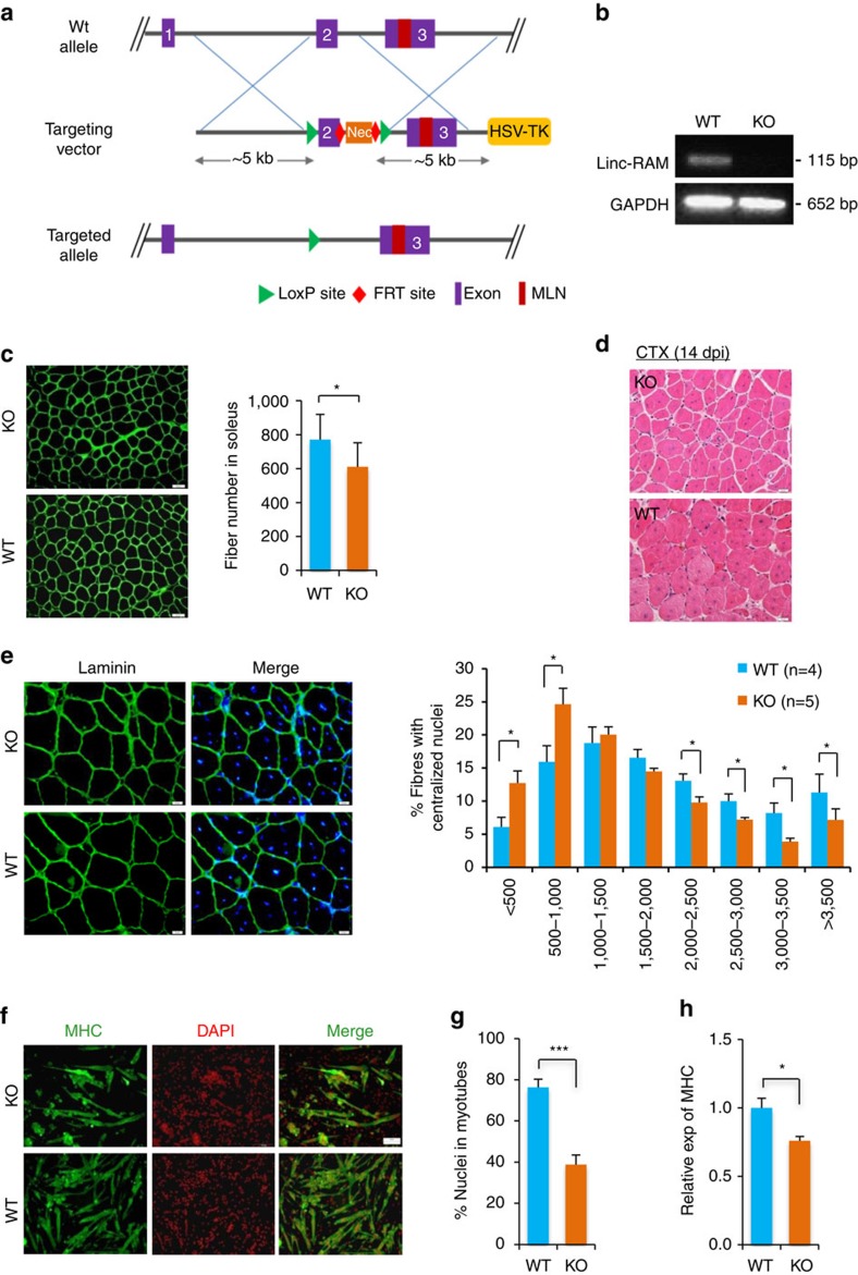
Figure 2. Linc-RAM knockout mice exhibits delayed muscle regeneration.
(a) Strategy for generation of Linc-RAM knockout mice. LoxP sequences were inserted in the flanking of exon 2 of the Linc-RAM gene. (b) The exon 2 deletion in knockout muscle was confirmed by RT–PCR. (c) The numbers of myofibers in soleus muscle of wild-type (WT; n=7) and knockout (KO) mice (n=9) were calculated based on laminin staining (left). Scale bars, 50 μm. (d) Representative hematoxylin and eosin (H&E)-stained sections of TA muscle 14 days post injury (14 dpi) induced by CTX injection. Scale bars, 50 μm. (e) Cross-sectional area of regenerated myofibers with centralized nuclei, calculated from the laminin-stained sections (left). Scale bars, 20 μm. (f) The differentiation of primary myoblasts isolated from 3-week-old Linc-RAM KO and WT littermates were analysed by inducing differentiation in DM for 36 h and staining with MHC. The presented images are representatives of three independent experiments. Scale bars, 50 μm. (g) Fusion index in f were calculated. (h) The expression of MHC in f was detected by RT–qPCR. GAPDH was the internal control. Values are means±s.e.m. The statistical significance of the difference between two means was calculated with the t-test, *P<0.05, ***P<0.001.

