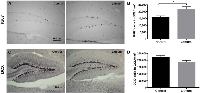
Figure 5. Lithium boosted hippocampal NSPC proliferation but not neuronal differentiation.
(A) Ki67 immunoreactivity in the granule cell layer (GCL) of control and lithium-treated animals. In each group n = 6. (B) Bar graph showing the quantification of Ki67+ cells in the SGZ after 28 days of lithium treatment. Significance was considered at p-values < 0.05. (C) DCX immunoreactivity in the granule cell layer (GCL) of control and lithium-treated animals. (D) Bar graph showing the quantification of DCX+ cells in the GCL after 28 days from the onset of lithium treatment. In each group n = 6. Data are presented as mean ± SEM.

