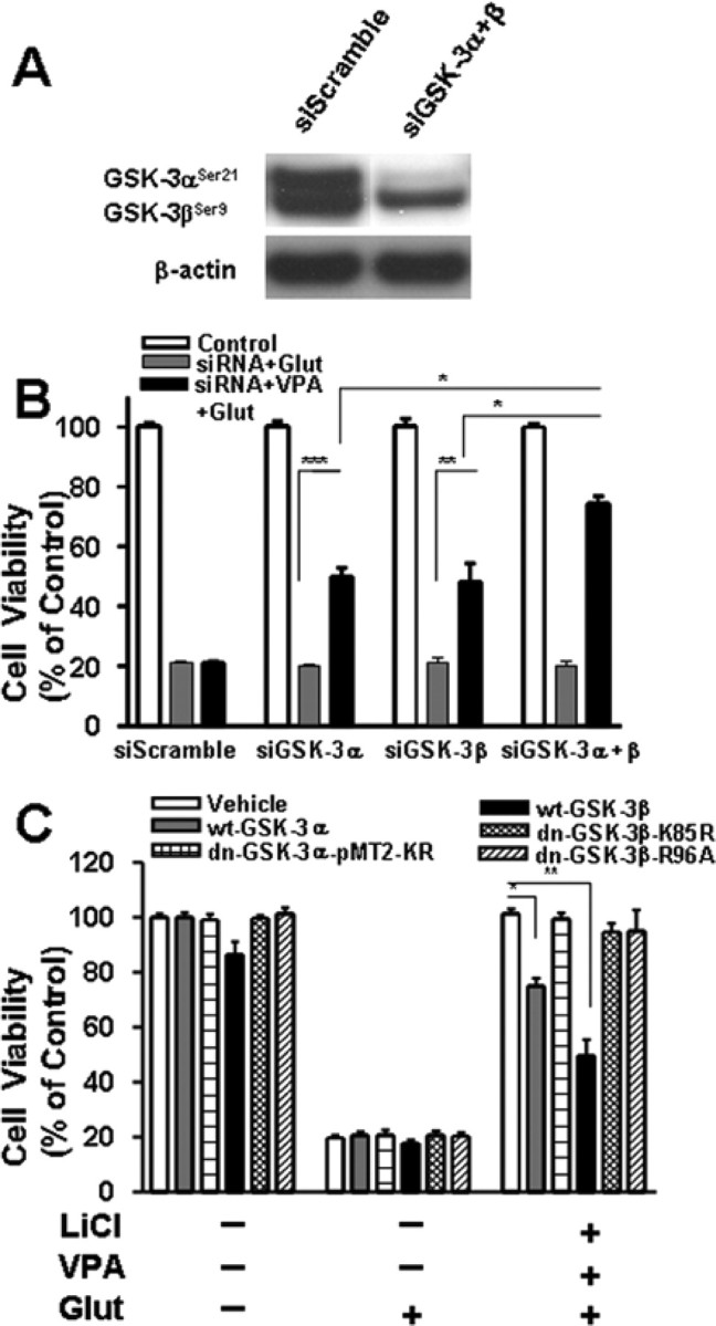
Figure 7.

Effects of rat GSK-3 isoform-specific siRNAs and GSK-3β plasmids on glutamate-induced excitotoxicity. Cells were transfected with 100 nm siRNA specific to either GSK-3α (siGSK-3α; αP1269) or GSK-3β (siGSK-3β; βP555) before plating by electroporation. Scrambled siRNA (siScramble) at a corresponding concentration was used as the transfection control. Western blotting revealed that protein levels of both GSK-3α and GSK-3β were markedly decreased 6 d after transfection with a mixture of siRNA for GSK-3α and siRNA for GSK-3β (siGSK-3α + β) (A). Transfected cells were also treated with 0.8 mm VPA or its vehicle from 6 to 12 DIV and then exposed to 50 μm glutamate (Glut) for 24 h (B). Cell viability was measured by MTT assay and expressed as means ± SEM of percentage of vehicle-treated control from three independent experiments. *p < 0.05, **p < 0.01, ***p < 0.001 between the indicated groups. Note that transfection with siGSK-3α and/or siGSK-3β was neuroprotective only in the VPA-treated groups. In another experiment (C), CGCs were transfected at the time of plating with a plasmid of wild-type GSK-3α (wt-GSK-3α), GSK-3α dominant-negative mutant (dn-GSK3α-pMT2-KR), wild-type GSK-3β (wt-GSK-3β), or GSK-3β dominant-negative mutant (dn-GSK-3β-K85R or dn-GSK-3β-R96A). Cells were then treated with a combination of LiCl (3 mm) and VPA (0.8 mm) or vehicle from 6 to 12 DIV, followed by 24 h exposure to glutamate (50 μm). Cell viability determined by MTT assay was expressed as means ± SEM from three independent experiments. Note that neuroprotection elicited by lithium and VPA was attenuated by wild-type GSK-3α or GSK-3β but not their dominant-negative mutants.
