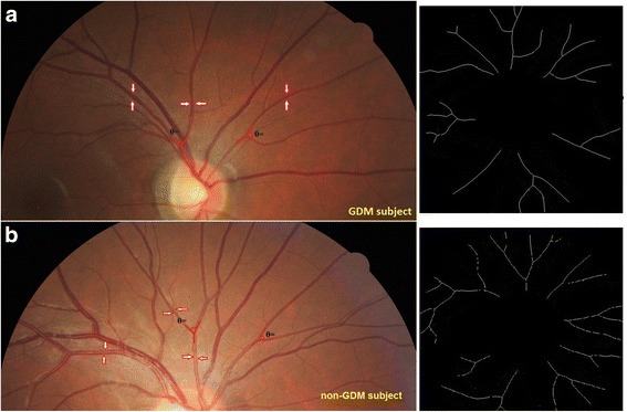
Fig. 2.

Comparison of retinal vasculature between a GDM (a) and a non-GDM GUSTO mother (b). Red arrows indicate retinal arterioles. Angle highlighted in red lines indicate retinal arteriolar branching angle. Black-and-white images on the right indicate retinal arteriolar fractal dimension. This GDM mother has narrower retinal arteriolar caliber (116.6 vs. 125.4 µm), narrower retinal arteriolar branching angle (1.26 vs. 1.31 Df), and lower retinal arteriolar fractal dimension (98.0 vs. 74.3 degrees) than the non-GDM mother
