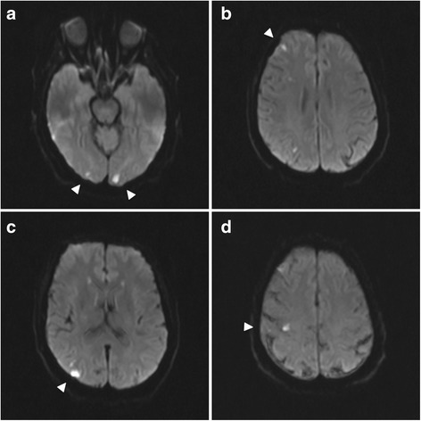
Fig. 1.

Magnetic resonance imaging of the brain. Scattered bilateral cerebral hemispheric small cortical-based acute infarcts (arrows) with a distribution suggestive of embolic phenomenon and alternatively watershed regions, including right occipital (a, c), left occipital (a), right frontal (b), and right parietal lobes (d)
