Fig. 3.

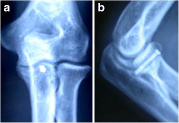
a X-ray of a 32-year-old male patient shows fracture of the ulnar coronoid process (Regan and Morrey type II). b Lateral X-ray 6 weeks after the treatment shows no displacement of the fracture

a X-ray of a 32-year-old male patient shows fracture of the ulnar coronoid process (Regan and Morrey type II). b Lateral X-ray 6 weeks after the treatment shows no displacement of the fracture