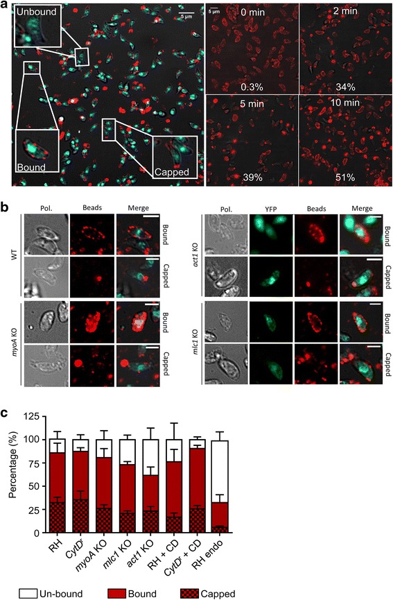
Fig. 10.

Analysis of bead translocation. a Three types of interactions of parasites with beads were observed (left panel); no interaction (unbound), beads located all around the parasite (bound), and beads translocated to the posterior end of the parasite (capped). Kinetics of bead translocation on wildtype parasites (RH) (0–10 min; right panel); the percentage of parasites showing a capped pattern at each time point is indicated. b Representative images of bead assays at 10 min in different genetic backgrounds, illustrating the binding and capping capacity of RH, myoA knockout (KO), act1 conditional knockout (cKO) and mlc1 cKO parasites. c The amount of bead binding and capping was quantified after 10 min incubation in different parasite strains: un-bound (white), bound (red) and capped (red/black patterned). c RH, Cytochalasin-D (CD) resistant (CytDr), myoA KO, mlc1 cKO, act1 cKO parasites, and RH incubated in Endo-buffer (RH Endo). Capping in the presence of 0.5 μM CD was also evaluated for wildtype and CytDr parasites. The percentage of bead interaction with the parasites’ surface was significantly reduced in the RH Endo and act1 cKO parasites. Capping still occurs in all KO mutant strains. Results are representative of at least three independent experiments ± standard deviation with at least 1000 parasites counted per experiment, per condition (n = 3)
