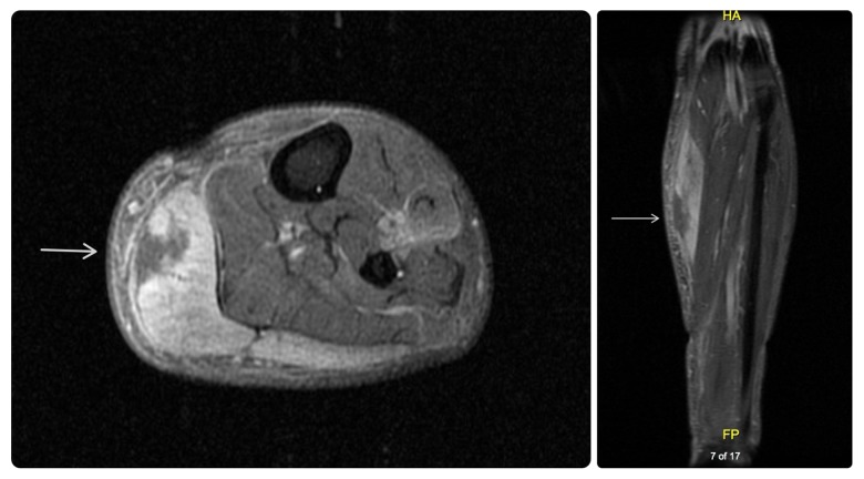
Figure 2.
Left calf MRI axial and coronal FSE T1 with contrast. MRI shows a 4.7×2.1×1.6 cm focus of non-enhancement (white arrow) in the inferior medial head of the gastrocnemius muscle and edema-like signal changes with enhancement of the rest of the gastrocnemius and extensor digitorum longus muscle, suggestive of diabetic myonecrosis.

