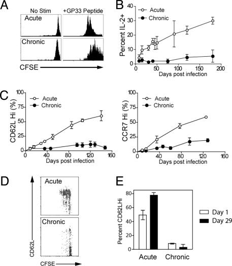
Fig. 4.
Impaired memory CD8 T cell differentiation during chronic infection. (A) CFSE-labeled acute memory (day 240) or chronic memory (day 240) CD8 T cells were stimulated in vitro with GP33 peptide, and division of Db/GP33+ CD8 T cells was assessed after 60 h. (B) The ability to produce IL-2 after in vitro stimulation with GP33 peptide was assessed at the indicated times by intracellular cytokine staining. Percentage indicates the percent of Db/GP33+ CD8 T cells from the spleen that produced IL-2; n = 2–7 for all points. The difference between acute and chronic infection is significant (P < 0.05) after day 8. (C) CD62L and CCR7 expression by Db/GP33+ CD8 T cells after acute versus chronic infection; n = 2–7 for all points. The difference between acute and chronic infection is significant (P < 0.05) after day 8 for CD62L and day 15 for CCR7. (D) Acute memory (day 100) and chronic memory (days 140–160) CD8 T cells were purified and transferred to naïve mice. Thirty-four days later, division and phenotype of CFSE-labeled, Db/GP33+ CD8 T cells and acute or chronic memory CD8 T cells were analyzed by costaining for CD62L. Results shown are for the spleen. Similar results were observed for lymph nodes, PBMC, and liver (data not shown). (E) CD62L expression on Db/GP33+ acute and chronic memory CD8 T cells in the PBMC after adoptive transfer to naïve recipients. There was a significant increase in the percentage of CD62LHi acute memory CD8 T cells (P < 0.01) after 34 days in naïve adoptive hosts, but no significant change in the percentage of CD62LHi chronic memory. Similar results were observed in the spleen (data not shown). n = two to three per group, and data are representative of four independent experiments. Similar results were observed for CCR7 and CD127 expression (data not shown). For all panels, similar results were observed for the Db/GP276+ CD8 T cells (data not shown).

