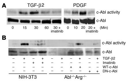
Figure 1.
TGF-β2 stimulates c-Abl kinase activity. (A) NIH-3T3 fibroblasts were grown to confluence, placed in 0.1% FBS DMEM overnight, and stimulated with 10 ng/ml TGF-β2 or 25 ng/ml PDGF-AB for the indicated times. Parallel plates were pretreated for 20 minutes with 10 μg/ml imatinib prior to growth factor addition. Following c-Abl immunoprecipitation, in vitro kinase activity was determined as described in Methods. Similar results were obtained with AKR-2B fibroblasts and TGF-β1 (data not shown). Lower half: Prior to immunoprecipitation and kinase assay, 100 μg of protein was used for c-Abl Western analysis. (B) NIH-3T3 cells, Abl–/–Arg–/– MEFs, or Abl–/–Arg–/– fibroblasts stably expressing (+) WT-c-Abl or dominant negative c-Abl (DN-c-Abl) were plated at 2.0 106 cells per 100-mm plate and grown overnight in 20% FBS DMEM. The medium was changed to 0.5% FBS DMEM for 24 hours, and the cultures were left untreated (–) or stimulated (+) with 10 ng/ml TGF-β2 for 30 minutes. Parallel plates were pretreated for 20 minutes with 10 μg/ml imatinib prior to growth factor addition. c-Abl kinase activity (top) and Western analysis (bottom) were determined as in A.

