Figure 1.
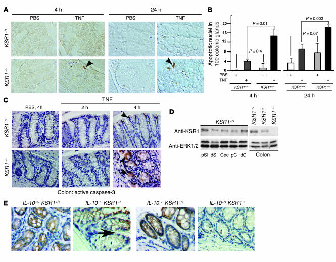
TNF induces apoptosis in KSR1–/– mouse colon epithelium in vivo. Mice were injected with TNF or PBS for the indicated times. Paraffin-embedded colon tissues were studied for apoptosis using ISOL staining. (A) Apoptotic nuclei labeled with peroxidase were visualized using DIC microscopy. Arrowheads indicate ISOL-labeled apoptotic nuclei. (B) The number of apoptotic nuclei found per 100 colonic glands. (C) Caspase-3 activity was determined by immunohistochemistry using anti–active caspase-3 antibody. Arrowheads indicate examples of caspase-3–positive cells detected by peroxidase. KSR1 expression in the gastrointestinal tract was determined by Western blot analysis of mucosal lysates (D) and immunohistochemistry (E). The arrow in E points to the transitional section of KSR1 expression in IL-10+/–KSR1+/– mouse colon. d, distal; p, proximal; SI, small intestine; Cec, cecum; C, colon. The data shown here are representative of 5 different experiments. Magnification, ×40.

