Figure 3.
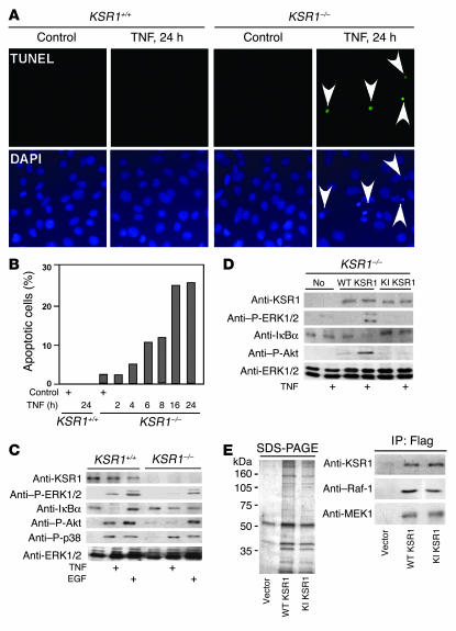
KSR1 regulates TNF-induced survival responses in MCE cell lines. (A) KSR1–/– MCE cells were treated with TNF for 24 hours and fixed for TUNEL and DAPI staining. Apoptotic cell nuclei in TUNEL staining were labeled with FITC (indicated by arrowheads) and visualized using fluorescence microscopy (magnification, ×40). (B) The percentage of cells undergoing apoptosis following TNF treatment for the indicated times is shown from a representative experiment. KSR1–/– MCE cells (C) or KSR1–/– MCE cells transiently transfected with WT KSR1 or KI KSR1 (D) were treated with TNF or EGF, and cellular lysates were prepared for Western blot analysis with the indicated antibodies. KSR1 and its coprecipitated proteins from KSR1–/– MCE cells transiently transfected with vector, WT KSR1 or KI KSR1 were separated by SDS-PAGE and stained with colloidal blue. Raf-1 and MEK bound to KSR1 was determined by Western blot analysis with anti–Raf-1 or anti-MEK1/2 antibodies (E). Shown are representative data from 3 separate experiments.

