Figure 1.
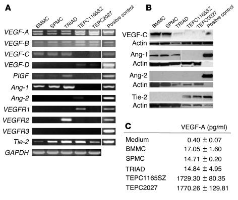
Expression of proangiogenic factors and receptors in mast cells and plasmacytoma cells. (A) Total RNA extracted from primary mast cells from bone marrow (BMMCs, cultured with IL-3; TRIAD cells, cultured with SCF, IL-6, and IL-10) and spleen (SPMCs, cultured with IL-3), and from plasmacytoma cell lines (TEPC1165SZ and TEPC2027), was subjected to RT-PCR using specific primer. Representative results are shown. (B) Western blot analysis of lysates from 2 × 10⁶ mast cells and plasmacytoma cells using specific antibodies. Loading accuracy was evaluated by reprobing with anti-actin antibodies. Positive controls: VEGF-C, lung tissue; Ang-1, recombinant Ang-1 (2.5 ng); Ang-2, recombinant Ang-2 (2.5 ng); Tie-2, human umbilical vein EC. (C) VEGF-A content in culture supernatants of mast cells and plasmacytoma cells, detected by specific ELISA. All cells were cultured at 106 cells/ml for 48 hours.

