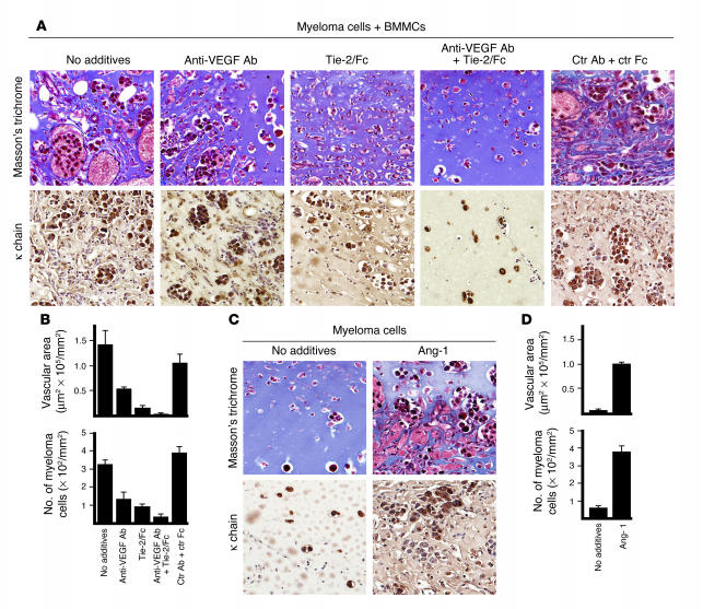
Figure 3.
Evidence supporting a contribution of mast cell–derived Ang-1 to neovascularization in vivo. Matrigel plugs (0.5 ml) containing plasmacytoma cells (TEPC2027 cells, 0.5 × 10⁶) alone or with BMMCs (0.5 × 10⁶) were inoculated s.c. into mice (3–5 mice per group) without additives or with anti–VEGF-A antibody (5 μg/ml), Tie-2/Fc (5 μg/ml), anti–VEGF-A antibody plus Tie-2/Fc (5 μg/ml each), control (Ctr) goat IgG plus control B7-1/Fc (5 μg/ml each), or recombinant Ang-1 (500 ng/ml). (A) Representative histological images of Matrigel plugs stained with Masson’s trichrome (top row), showing different degrees of neovascularization; and immunostained for κ light chain (bottom row), showing differing degrees of plasmacytoma cell infiltration under the experimental conditions tested. Original magnification, ×20. (B) Quantitative analysis (as described in the legend to Figure 2) of Matrigel neovascularization (top) and plasmacytoma cell infiltration (bottom) under the experimental conditions tested (3–5 plugs per group; 1 plug per mouse). (C) Representative microscopic images of Matrigel plugs containing plasmacytoma cells alone or with recombinant Ang-1, stained with Masson’s trichrome (top row) or immunostained for κ chain (bottom row), showing differing degrees of neovascularization and plasmacytoma cell infiltration. (D) Quantitative analysis (as described in the legend to Figure 2) of Matrigel neovascularization (top) and plasmacytoma cell infiltration (bottom) in the presence of plasmacytoma cells alone or with recombinant Ang-1 (3–5 plugs per group; 1 plug per mouse).

