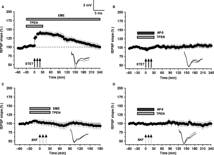
Figure 5.

Augmentation of L‐LTP and restoration of DA‐LTP in aged rat slices following Zn2+ chelation depends on NMDAR activity and protein synthesis. (A) STET coupled with co‐application of TPEN (5 μm) and emetine (EME, 20 μm) resulted in a potentiation that remained significant for 145 min (n = 12; P = 0.042) and decayed to baseline afterward. (B) STET failed to induce potentiation in the presence of NMDAR antagonist AP5 (50 μm) and TPEN (5 μm). The fEPSP response throughout the recording period was not significantly different from that of baseline (P > 0.05, n = 8). (C) SKF application coupled with application of TPEN (5 μm) and emetine (20 μm) completely suppressed the DA‐LTP (Wilcoxon's test, n = 6; P > 0.05). (D) SKF application failed to induce potentiation in the presence of NMDAR antagonist AP5 (50 μm) and TPEN (5 μm). The fEPSP response at the end of 4 h was not significantly different from that of baseline values (Wilcoxon's test, P > 0.05, n = 9). Error bars in all graphs represent ±SEM. Insets show representative fEPSP traces recorded at baseline (black solid line), 30 min after the respective tetanization/stimulation (dotted line), and at 240 min (gray solid line). Symbols as in Fig. 1. Scale bar for the traces 3 mV per 5 ms.
