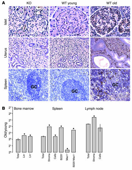
Figure 2.
p16INK4a expression in specific compartments by immunohistochemistry and cell purification. (A) Immunoperoxidase staining performed on paraffin-embedded sections of germ-line p16INK4a-deficient (KO), WT young (3.5 months), and WT old (25 months) murine tissues using an anti-p16INK4a antibody. Positively staining cells demonstrate both nuclear and cytoplasmic expression. GC, germinal center. (B) Relative expression ratios (old/young, log2 scale) of p16INK4a in specific compartments (average purity >94% for all fractions) of bone marrow (lin–, 2%; lin+, 97%), spleen (B220+, 48%; Mac1+, 9%; B220–Mac1–, 22%), and lymph node. Asterisks indicate that p16INK4a expression was undetectable in these cell populations from young mice, and therefore a minimum estimate of the fold increase is shown.

