
Figure 3.
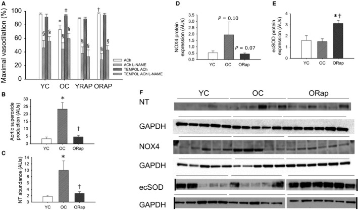
Oxidative stress in arteries of young and old untreated and rapamycin‐treated mice. (A) Maximal vasodilation to acetylcholine (ACh) in the absence or presence of the nitric oxide synthase inhibitor, L‐NAME, and the superoxide scavenger, TEMPOL, in carotid arteries from young untreated (N = 6), young rapamycin (Rap)‐treated (N = 6), old (OC) (N = 6) untreated and old Rap‐treated mice (N = 6). * denotes difference from young ACh alone, †denotes difference from Old ACh alone, ‡ denotes difference after TEMPOL from old ACh alone, and § denotes difference with L‐NAME from paired ACh response. Differences were assessed by repeated‐measures ANOVA with LSD post hoc. (B) Superoxide production measured by electron paramagnetic resonance in aortas from young and old untreated and old Rap‐treated mice (N = 6–9/group). (C) Abundance of nitrotyrosine (NT), a marker of oxidative stress, in aortas from young and old untreated and old Rap mice (N = 5–6/group). (D) NADPH oxidase 4 (NOX4) expression in aortas from young and old untreated and old Rap mice (N = 5‐6/group). P = 0.1 vs. young, P = 0.07 vs. old. (E) Extracellular superoxide dismutase (ecSOD) expression in aortas from young and old untreated and old Rap mice (N = 5–6/group). (F) Blot images for NT, NOX4, ecSOD, and the associated normalizer, GAPDH. Black vertical lines indicate where blot image was digitally cut to reposition groups in the same order as the summary graphs, where applicable. Expression is presented as a ratio to GAPDH to account for differences in protein loading. Differences were assessed by one‐way ANOVA with LSD post hoc. For B–E, * denotes difference from young untreated and † denotes difference after old untreated. Data are means ± SEM, P ≤ 0.05