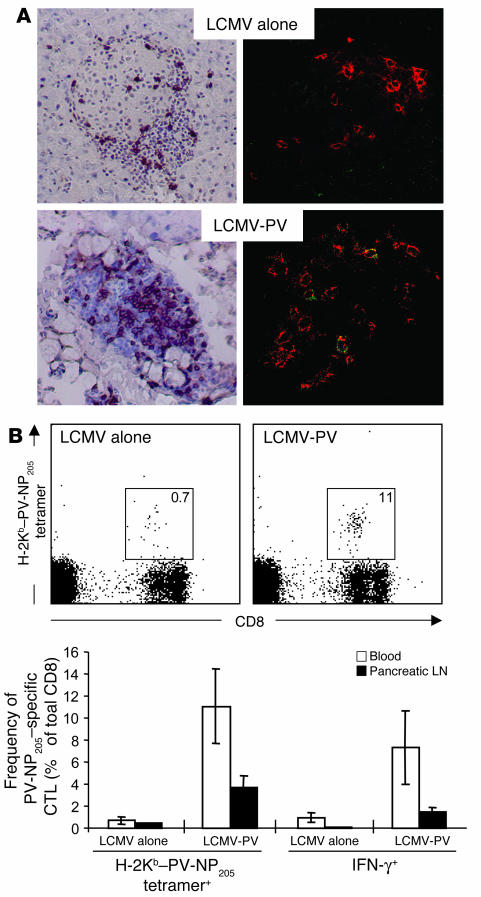
Figure 3.
Sequential infection with LCMV and PV results in accumulation of PV-NP205–specific CD8 T cells in the islets of Langerhans. (A) RIP-NP mice were infected with 105 PFU LCMV. After 4 weeks, one group of mice received a secondary infection of PV (105 PFU, i.p.). Left panels, pancreata were harvested at week 3 after secondary infection and 6-μm tissue sections were stained for cellular infiltration with a monoclonal antibody against CD8. Sections were counterstained with hematoxylin. Right panels, pancreata were harvested at day 5 after secondary infection and 6-μm tissue sections were cut and were stained for CD8 T cells with rhodamine X–conjugated anti-CD8 (red) and for PV-NP205–specific CD8 T cells with allophycocyanin-conjugated H-2Kb–PV-NP205 tetramers (green). Note that only after sequential infection with LCMV followed by PV are PV-NP205–specific CD8 T lymphocytes (yellow) found in the islets of Langerhans. Original magnification, ×20. (B) Expansion of PV-NP205–specific CD8 T cell populations in blood and pancreatic lymph nodes after secondary PV infection. Upper panels, flow cytometry of PV-NP205–specific CD8 T cells in the blood of LCMV-immune mice that did or did not receive secondary infection with PV, as detected by H-2Kb–PV-NP205 tetramers; mean frequencies are indicated in boxed areas. Lower panel, frequencies of PV-NP205–specific CD8 T cells were determined by flow cytometry using H-2Kb–PV-NP205 tetramer staining and by ICCS for IFN-γ expression after 5 hours of in vitro stimulation with PV-NP205 peptide.

