
Figure 3. IL-17C deficient Apoe−/− mice display diminished aortic leukocyte, myeloid cell, macrophage and neutrophil content.
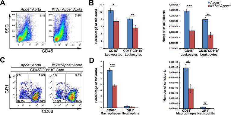
(A–D) 12 week WD-fed Apoe−/− and Il17c−/−Apoe−/− aortas with surrounding aortic adventitia were stained with anti CD45, CD11b, CD68, and Gr1 antibodies and assessed by flow cytometry. (A) Representative aortic CD45 staining and quantification (B) of 12 week WD Apoe−/− (blue) and Il17c−/−Apoe−/− (red) aortic CD45+ leukocyte, and CD45+CD11b+ myeloid cell content. (C) Representative CD68 and Gr1 staining within CD45+CD11b+ gated aortas. (D) Quantification of the percentage and number of CD68+ macrophages and Gr1+ neutrophils per Apoe−/− (blue) and Il17c−/−Apoe−/− (red) aorta. n= 5 Apoe−/− mice, n=8 Il17c−/−Apoe−/− mice, 3 independent experiments. Means±SEM are shown, * - p<0.05, ** - p<0.01, *** - p<0.001.