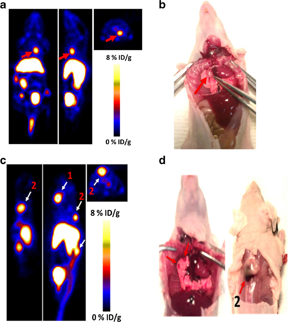
Fig. 6.
a Coronal (left), sagittal (middle), and axial (left) PET images of metastatic model of CHO-CXCR4 tumors at 1 h post-injection of Al[18F]NOTA-T140 with a %ID/g of 9.33, calculated from the PET images. Red arrow represents lesion location. b Photograph of a, tumor location shown with red arrow. c Mouse bearing two CXCR4-positive metastatic tumors on the spine, with %ID/g of 12.2 (for 1) and 9.8 (for 2), at 1 h post-injection of Al[18F]NOTA-T140. d Ventral (left) and dorsal (right) photograph of c. Tumor locations are shown with white arrows.

