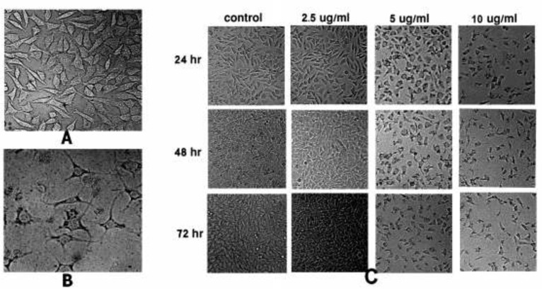
Figure 1.
Morphological alterations of HepG2 cells after exposure to different concentrations of SVT, observed by normal inverted light microscopy. (A) Untreated cells. (B) Morphological changes in HepG2 cells after 24 h treatment with 10 µg/mL of SVT. Detachment of cells from the dish, cell rounding, cytoplasmic blebbing, chromatin condensation and irregularity in shape are observable. (C) Significant decrease in attached cell population after treatment with different concentrations of SVT for various time periods

